Unique Registration Number: GSInC-I-000303
Innovator Name:
- Aakash Koli
- Pushpam Mandrekar
- Fraser Silveira
Name of a Mentor:- Prof. Pratiksha Shetgaonkar
Name of School/College/Startup/Organisation: Shree Rayeshwar Institute Of Engineering And Information Technology, Shiroda-Goa.
Contact No: +917066395194
Contact Email: koliaakash42@gmail.com
Project Objective:
- Develop a sophisticated computational tool using deep learning for automated segmentation and categorization of brain tumors in medical images.
Abstract:
- Accurate segmentation of brain tumors is crucial for diagnosis and treatment planning in neuroimaging. Leveraging semi-supervised learning approaches can potentially enhance segmentation performance by utilizing both labeled and unlabeled data. In this paper, we propose a novel methodology for semi-supervised brain tumor segmentation, employing context restoration for pseudo label generation as a pretext task. We investigate the efficacy of this approach across three prominent variants of the UNet architecture: the standard UNet, Attention UNet, and UNet++. By varying the proportion of labeled and unlabeled data, we conduct a comprehensive comparative analysis to evaluate the segmentation performance of each model. Our results demonstrate the effectiveness of the proposed method in enhancing segmentation accuracy, particularly in scenarios with limited labeled data. Furthermore, we provide insights into the relative performance of different UNet variants under varying degrees of data availability, offering valuable guidance for practitioners in selecting an appropriate model for semi-supervised brain tumor segmentation tasks.
Project Outcome/result/findings:
I. EXPERIMENTAL RESULTS
The proposed methodology focuses on developing semi-supervised segmentation models with a context restoration task for brain tumor detection. The architecture involves multiple steps from pre-processing to model deployment. In Fig. 1, the suggested architecture is displayed, illustrating the workflow. To facilitate this process, we utilize the Google Colab platform, a cloud-based environment. The platform is equipped with T4, and P100 GPUs, along with a 16 GB RAM runtime, optimizing the training of machine learning models.
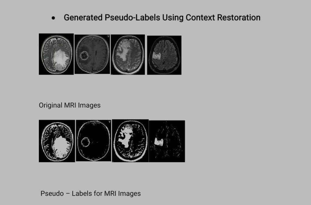
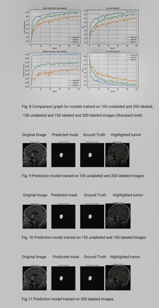
We conducted extensive experiments on the Colab platform, training each model for 100 epochs with datasets comprising varying proportions of unlabeled and labeled data. For the 33.33% unlabeled and 66.66% labeled model, we utilized 100 unlabeled images along with 200 labeled images. Similarly, for the 50% unlabeled and 50% labeled model, we employed 150 labeled images and 150 unlabeled images. Finally, for the 100% labeled data model, we utilized 300 labeled images. Additionally, prior to these experiments, we trained a context restoration model using 1000 tumorous images and 500 non-tumorous images. Through these rigorous experiments, we aimed to investigate the influence of dataset size and label availability on the performance and robustness of each model in brain tumor segmentation tasks.
Moreover, we utilized an additional 100 images for generating pseudo labels, which were not included in the training set.

Innovative Approach: